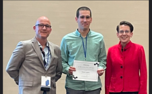
Many congratulations to Abdul on the award of his PhD! His thesis is entitled Characterisation of Battery Materials Using Surface Science Techniques and describes novel methods and results using ToF-SIMS and HAXPES to explore the interfacial chemistry of electrode materials. Thanks to his External Examiner Prof Sven Schroeder from the University of Leeds. Abdul is currently Lecturer at the University of Jeddah. Well done Abdul!
Publication – Visualizing fungicide mobility in tomato leaves using DESI
Another publication for Akhila, resulting from our collaboration with Syngenta, Waters Corp. and colleagues in the Manchester Institute of Biotechnology and The Department of Earth and Environmental Sciences:
DOI: 10.1039/D4AN01309C (Paper) Analyst, 2024, Advance Article 
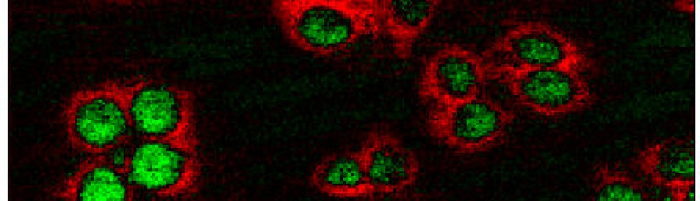
Publication – Multi-Modal Brain Bioimaging
Our collaboration with the Medicines Discovery Catapult led by Irma results in a publication in a Special Issue of the International Journal of Molecular Sciences – Molecular Advances in Analytical Techniques for Biological and Medical Research: 2nd Edition
The paper is entitled Accumulation of Bioactive Lipid Species in LPS-Induced Neuroinflammation Models Analysed with Multi-Modal Mass Spectrometry Imaging
Int. J. Mol. Sci. 2024, 25(22), 12032; https://doi.org/10.3390/ijms252212032

SIMS-24 Conference

It was a busy week at SIMS-24 in La Rochelle, France, with presentations from Abdul, Akhila, Matija, Alex and Nick. Congratulations to the Co-Chairs Alain Brunelle, Jean-Paul Barnes and their teams for a great conference!
Publication – GCIB developments
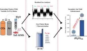
A study from Matija’s PhD on the application of Reactive Gas Cluster Ion Beams has been published in Analytical Chemistry.
Reactive Gas Cluster Ion Beams for Enhanced Drug Analysis by Secondary Ion Mass Spectrometry
Matija Lagator, Bilal Patel, Sadia Sheraz, and Nicholas Lockyer
Analytical Chemistry Article ASAP
DOI: 10.1021/acs.analchem.4c02144
Nature Reviews Publication
Together with fellow SIMS experts from around the world we have published a new Primer ‘Secondary Ion Mass Spectrometry‘ in Nature Reviews Methods. The manuscript describes the operating principles of SIMS and outlines how the instrument geometry and operational parameters enable different modes of operation and information to be obtained. Applications, including materials science, surface science, electronic devices, geosciences and life sciences, are explored, finishing with an outlook for the technique.
You can view the formatted article here
PhD for Matija
Many congratulations to Matija for the award of his PhD on the ‘Development of Novel High Energy Cluster Ion Beam Methodology for Molecular Analysis and Imaging‘. Thanks to the external examiner Prof Mel Bailey from The University of Surrey and internal examiner Dr Drupad Trivedi. Matija will be joining the SIMS group led by Dr Felicia Green at the Rosalind Franklin Institute. Well done Matija and good luck!
Award for Akhila
Many congratulations to Akhila for winning the Peter Ryan Award for the best presentation at the BMSS Ambient Ionisation meeting in Birmingham! The title of Akhila’s oral presentation was titled ‘Visualizing Fungicide Mobility in Tomato Leaves with DESI Mass Spectrometry Imaging‘. The Award was sponsored by KR Analytical. Well done Akhila!
New Collaboration: Multimodal Ion Beam Imaging facility
We are excited to announce a new collaboration with The Surrey Ion Beam Centre on a new £3m project ‘Multimodal 3D elemental and molecular imaging at the sub-micron scale‘ funded by the EPSRC. The project, led by Prof Melanie Bailey at the University of Surrey, will be achieved.by combining MeV ion beam analysis (X rays, gamma rays, backscattered particles) and water cluster Secondary Ion Mass Spectrometry (SIMS) for the first time in a single instrument.
Happy 80th John!
We recently got together with some alumni and friends of the group to celebrate John Vickerman’s 80th Birthday with a meal and drinks in Manchester city centre. It was great to catch up with everyone. Congratulations John!
Publication – Nanoscale Advanced Materials Engineering
Our collaboration with Prof Richard Curry results in a publication in the journal Advanced Engineering Materials:
A High-Resolution Versatile Focused Ion Implantation Platform for Nanoscale Engineering
Adv. Eng. Mater., 25: 2300889. https://doi.org/10.1002/adem.202300889
Publication – Data processing
We have co-authored the following paper led by Prof Satoka Aoyagi and her group at Seikei University, Japan.
Anal. Chem. 2023, 95, 40, 15078–15085

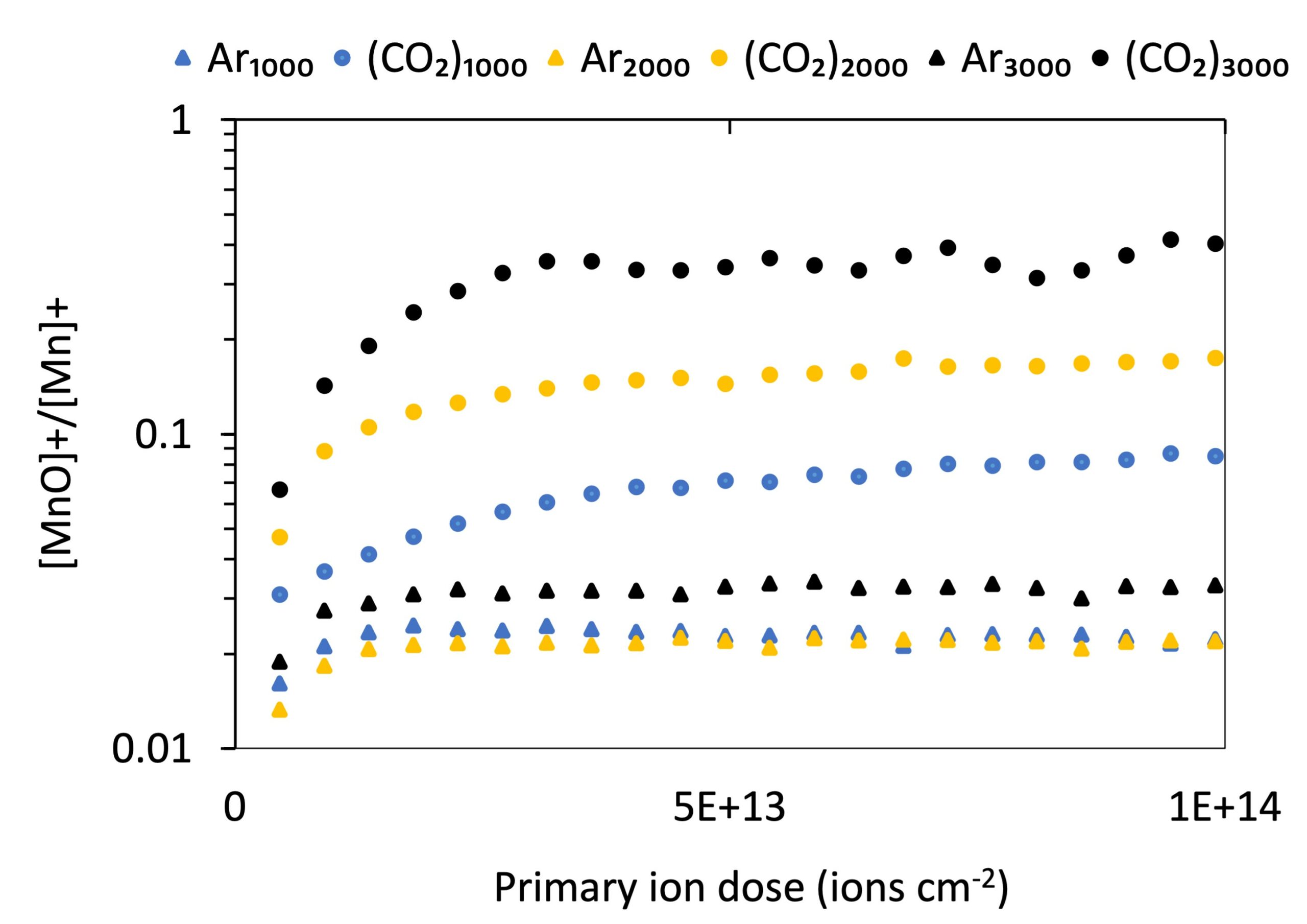
Publication – GCIBs for metal oxide analysis
Congratulations Abdul on your recent publication:
Secondary ion mass spectrometry analysis of metal oxides using 70 keV argon, carbon dioxide, and water gas cluster ion beams
Journal of Vacuum Science & Technology B 41, 044007 (2023)


Fully-funded PhD studentships available
Search ‘Lockyer’ on FindaPhD.com to see current funded PhD opportunities:
We are currently recruiting to the following positions:
(BBSRC DTP) Shedding new light on the spatial lipidomics of disease progression using vibrational spectroscopy and mass spectrometry
POSITION FILLED
Matija awarded for SIMS 23 presentation
Congratulations Matija on winning the Rowland Hill Award for best student presentation at SIMS 23 in Minneapolis! Matija’s award-winning presentation was entitled ‘“Effects of Reactive Gas Cluster Ion Beams on Yields and Matrix Effects in SIMS”