Three-dimensional mass spectral imaging of HeLa-M cells – sample preparation, data interpretation and visualisation.
John S. Fletcher, Sadia Rabbani, Alex Henderson, Nicholas P. Lockyer, John C. Vickerman
Rapid Commun. Mass Spectrom. 25 (2011) 925-932
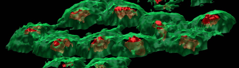
Topography corrected, 3D visualisation of the membrane (green, m/z 184.1, phosphocholine) and nucleus (purple, negative scores on principal component 5) chemistry in the fixed and freeze‐dried HeLa‐M cells following the use of PCA to identify the interface between the cells and the substrate to allow more representative data reconstruction. The visualised area is the same as the analysis area in the SIMS experiment, 250 × 250 μm2, but inclined in order to aid 3D visualisation. Video rendering by Alex Henderson